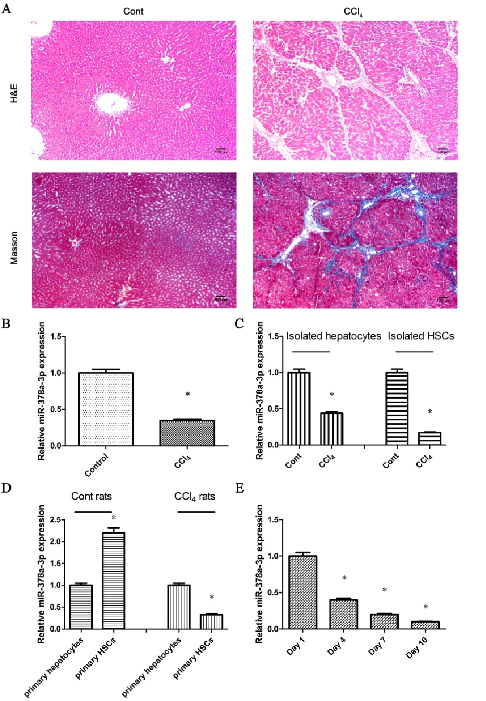
Fig. 1. miR-378a-3p expression in CCl4-treated hepatic fibrotic tissues and primary HSCs. (A) Hepatic fibrotic and control tissues (n=6) were detected by H&E staining (×100) and Masson staining (×100), respectively. (B) miR-378a-3p expression was detected in fibrotic liver tissues and control liver tissues (n=6). (C) Compared with healthy controls, miR-378a-3p expression was decreased in HSCs and hepatocytes isolated from CCl4-rats. (D) In healthy rats, miR-378a-3p expression was significantly higher in primary HSCs compared with that in primary hepatocytes. After CCl4 treatment, miR-378a-3p expression was reduced in primary HSCs compared with primary hepatocytes. (E) miR-378a-3p expression was detected in primary HSCs during different culture days. Each value is the mean ± SD of three experiments. *P<0.05 compared with the control or primary hepatocytes.